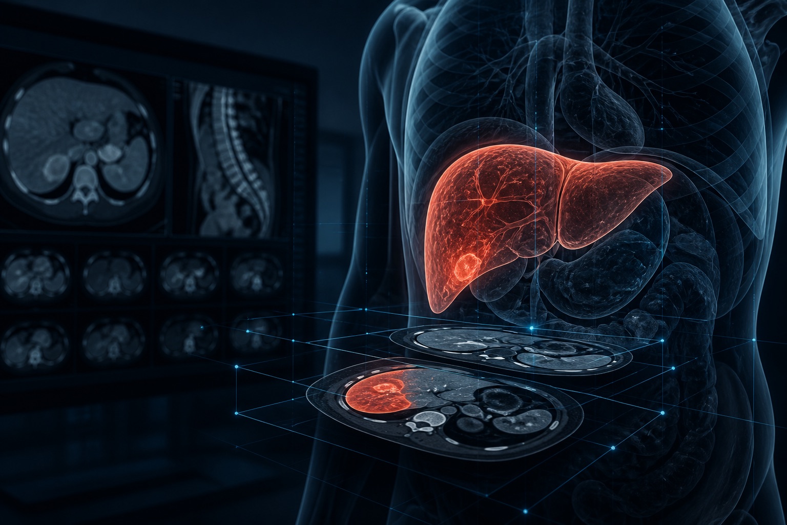
On August 24, 2026, Beijing time, QbitAI’s public feed highlighted research on LiON for diagnosing liver malignancies; the related paper had been published in Nature Medicine on August 19. LiON, short for Liver DiagnOsis Network, is an AI system that processes contrast-enhanced CT images. The study integrated it into the existing clinical workflow as an additional review step alongside physicians, and did not describe a process in which the system makes clinical diagnoses without them.
Research institutions and system structure
The work was conducted by researchers from Shengjing Hospital of China Medical University, Alibaba’s DAMO Academy, the First Affiliated Hospital of Zhejiang University School of Medicine, and other institutions. Participating organizations span several Chinese cities and include collaborators in France and the United Kingdom. The paper identifies missed or delayed diagnoses in high-volume radiology workflows as the clinical problem and evaluates a diagnostic safety net that can fit into routine practice. ClinicalTrials.gov lists Shengjing Hospital as the sponsor organization for the single-arm study and Yu Shi as principal investigator.
LiON processes multiphase contrast-enhanced CT. Noncontrast, arterial, and venous phases are required, while a delayed phase can be added when available. The paper’s public extended-figure caption describes four components—an image encoder, backbone, classifier, and segmenter—that use multiscale features for end-to-end processing. The system produces a patient-level malignancy risk score and segments eight liver-lesion types. Its classifier and segmenter use learnable queries and dense cross-attention; when clinical features are available, they can be integrated into patient-level predictions through two-stage score-based logistic regression.
Retrospective validation and subgroup results
The researchers trained LiON on data from 6,443 patients, then retrospectively validated it across 22,251 patients in multicenter and real-world cohorts. The paper reports an overall AUC of 0.975 for malignancy diagnosis, with a 95% confidence interval of 0.971–0.979. AUC measures a model’s ability to distinguish positive from negative cases; it is not the percentage of all cases classified correctly. The 0.975 figure therefore cannot be restated as 97.5% accuracy, and it applies only to the study’s specified data and evaluation methods.
The paper also reports results for two underlying liver-disease subgroups. Among patients with hepatic steatosis, the malignancy-diagnosis AUC was 0.971, with a 95% confidence interval of 0.952–0.985. Among patients with cirrhosis, it was 0.924, with a 95% confidence interval of 0.901–0.946. These figures describe performance in the study samples; the available evidence does not establish equivalent performance across every hospital, scanner, or healthcare system.
Single-arm trial and collaborative workflow
The team then conducted a single-arm interventional study at Shengjing Hospital in Shenyang, enrolling 10,333 patients under registration NCT07153783. The registry states that the study began on September 1, 2025, reached primary completion on October 29, and ended on November 7. It was neither randomized nor blinded and had no control arm. The prespecified primary endpoint required the lower bound of the 95% confidence interval for the malignancy-diagnosis AUC to exceed 0.900. The paper reports an AUC of 0.952, with a 95% confidence interval of 0.942–0.961, meeting that statistical threshold.
Within the clinical workflow, LiON operated as an additional reader alongside physicians. The paper’s abstract reports that the collaborative process identified 51 lesions omitted from the initial reports and confirmed on subsequent review; 15 were malignant. It also led to 37 amended radiology reports, 22 escalations to multidisciplinary teams, and changes in clinical management for some patients. These are secondary outcomes from a single-arm study. Without a randomized controlled comparison, the numbers alone cannot establish the net clinical benefit over standard practice.
Media representation and research boundaries
QbitAI’s feed headline described LiON as a liver-cancer AI model and said it could identify one-centimeter tumors; the short feed summary only confirms that DAMO Academy, Shengjing Hospital, and other institutions developed the system. The paper covers the broader category of liver malignancies, and neither its public abstract nor the trial registry reports one centimeter as a detection limit, average lesion size, or stratified result. This report therefore does not reduce every case to primary liver cancer or use a lesion-size figure that those public materials do not verify.
The authors state that prospective comparative studies across different healthcare systems are still needed to assess effects on clinical outcomes. Patient-level data are not public because of ethical and privacy restrictions; qualified researchers must seek access through governance review, ethics approval, and a data-use agreement. The complete LiON code is also unavailable because of proprietary infrastructure dependencies and pending patent protection, while the public repository contains only key algorithmic components and inference logic. Alibaba has filed related patent applications, and four authors disclosed that they are Alibaba employees who own company stock.